Intravascular Ultrasound
What is Intravascular Ultrasound (IVUS)?
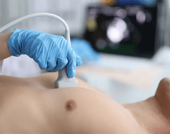
Intravascular Ultrasound (IVUS) is an advanced, catheter-based imaging technique that allows doctors to view detailed cross-sectional images of blood vessels from the inside. Unlike traditional ultrasound, which is performed from outside the body, IVUS uses a tiny ultrasound probe attached to a catheter inserted directly into the blood vessel.
This high-resolution technology helps assess arterial blockages, vessel size, plaque buildup, and overall vascular health with precision.
When is IVUS Used?
IVUS is most commonly used in patients with:
- Peripheral Artery Disease (PAD)
- Chronic leg pain or claudication
- Non-healing ulcers or wounds on the feet/legs
- Suspected arterial narrowing or blockages
- Prior abnormal imaging (angiogram, CT, or Doppler studies)
- Evaluation before or after stent placement or angioplasty
Why Choose IVUS Over Other Imaging?
IVUS provides real-time, high-definition imaging from inside the artery, offering several advantages:
- More accurate measurement of vessel diameter and plaque size
- Visual confirmation of blockages and stenosis severity
- Helps guide stent placement and ensures complete opening of treated arteries
- Identifies hidden or complex lesions that may be missed by traditional imaging
How the Procedure Works
- A catheter with an ultrasound probe at its tip is inserted into the artery (usually in the leg or groin).
- The catheter is guided through the blood vessel using X-ray or fluoroscopy.
- The ultrasound probe rotates inside the artery, capturing real-time cross-sectional images.
- The data is analyzed immediately to determine treatment options or confirm results of an intervention.
IVUS is often performed alongside a Lower Extremity Arteriogram or during an angioplasty or stent procedure.
Benefits of IVUS
- Provides unmatched precision for diagnosis and treatment planning
- Reduces the chance of under- or over-treatment
- Can prevent complications by optimizing device placement
- Painless and performed under local anesthesia with sedation
- Allows personalized vascular care tailored to the patient’s exact anatomy
What to Expect
- IVUS is typically done in a hospital or specialized vascular lab
- Procedure time is usually 30–60 minutes
- Most patients go home the same day after a short observation period
- Walking and daily activities can resume within 24 hours in most cases
Risks and Safety
IVUS is considered very safe when performed by a trained vascular specialist. Potential (rare) risks may include:
- Minor bleeding or bruising at the catheter insertion site
- Temporary discomfort
- Infection or allergic reaction (extremely rare)
Dr. Suri and his team follow strict safety protocols to ensure patient well-being at every step.